Systematic vs. targeted MRI/ultrasound fusion prostate biopsy among men with visible lesions

©Dan Oldfield
In men with PI-RADS 3 to 5 category lesions on their prostate MRI scans, Patel et al.
demonstrate improved detection of clinically significant cancer (as well as a small decrease in
detection of clinically insignificant disease) when targeted biopsies are added to standard
systematic biopsies.
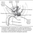
With a targeted biopsy, MRIs of the suspected cancer are fused with real-time ultrasound
images, creating a map of the prostate that enables doctors target the suspicious areas.
With transperineal biopsies the needle passes through the perineum, rather than the rectum.
The analyses revealed that adding biopsies targeting the MRI-visible lesions improves the
detection of clinically significant (≥GG2) cancer relative to a 12-core systematic transrectal
biopsy or a 20-core systematic transperineal biopsy. Adding targeted biopsies did not appear to
meaningfully increase detection of clinically insignificant disease.
While targeting lesions in men with positive MRI results appears to improve the yield of the
biopsy session, an important follow-up question is whether cognitive targeting provides
diagnostic yield similar to fusion biopsy platforms used at most large academic centers.
Urology. Jan 11, 2022 Practice Update